Saturday, February 25, 2023

The patient monitor


Patient monitor commonly used in hospitals is a medical device that continuously monitors various physiological parameters of a patient's health status, such as heart rate, blood pressure, respiratory rate, oxygen saturation, and temperature.

There are several types of patient monitors available, but most hospital monitors have a central display unit that shows real-time readings of the patient's vital signs. The display unit may also have alarms that alert healthcare professionals when the patient's vital signs fall outside of a predetermined range.

Patient monitors may also have the capability to store data over time, allowing healthcare professionals to review trends and changes in a patient's health status. Some patient monitors may also include additional features, such as the ability to monitor electrocardiogram (ECG) readings, invasive blood pressure monitoring, and capnography (monitoring of carbon dioxide levels in exhaled air).

Overall, patient monitors play a critical role in monitoring the health status of patients in hospitals, allowing healthcare professionals to quickly detect and respond to changes in a patient's vital signs.

Patient monitors are typically used in hospitals whenever a patient needs continuous monitoring of their vital signs. This includes patients who are:

  1. Undergoing surgery: During surgery, patient monitors are used to monitor the patient's vital signs and ensure that they remain stable throughout the procedure.

  2. In critical care units: Patients in critical care units, such as intensive care units (ICUs), may require continuous monitoring of their vital signs due to the severity of their illness or injury.

  3. Recovering from anesthesia: After surgery, patients may be monitored with a patient monitor while they recover from the effects of anesthesia.

  4. Receiving medication: Patients receiving certain medications, such as those that affect the heart or respiratory system, may require continuous monitoring to ensure their safety and efficacy.

  5. Experiencing a medical emergency: Patients experiencing a medical emergency, such as a heart attack or stroke, may require immediate and continuous monitoring of their vital signs.

Overall, patient monitors are an important tool in hospital settings, allowing healthcare professionals to continuously monitor a patient's vital signs and quickly detect any changes that may require intervention.

  Physiological parameters that can measure using a patient monitor

A patient monitor can measure several physiological parameters of a patient's health status, including:

  1. Heart rate: This parameter measures the number of heartbeats per minute and is typically measured using electrocardiogram (ECG) leads placed on the patient's chest.

  2. Blood pressure: Blood pressure can be measured non-invasively using a blood pressure cuff, or invasively using a catheter inserted into an artery.

  3. Respiratory rate: This parameter measures the number of breaths a patient takes per minute and is typically measured using sensors placed on the patient's chest or abdomen.

  4. Oxygen saturation (SpO2): This parameter measures the percentage of oxygen saturation in the blood and is typically measured using a pulse oximeter that clips onto the patient's finger or earlobe.

  5. Temperature: This parameter measures the patient's body temperature and is typically measured using a thermometer inserted into the patient's mouth, ear, or rectum.

  6. Carbon dioxide levels (EtCO2): This parameter measures the amount of carbon dioxide in exhaled air and is typically measured using a capnograph sensor placed on the patient's airway.

  7. Electrocardiogram (ECG): This parameter measures the electrical activity of the heart and is typically measured using ECG leads placed on the patient's chest.

  8. Invasive blood pressure: This parameter measures the patient's blood pressure using a catheter inserted into an artery.

These parameters are commonly measured by patient monitors in hospitals, allowing healthcare professionals to continuously monitor a patient's health status and quickly detect any changes that may require intervention.

The main functional blocks that are commonly used in patient monitors are:

  1. Sensors and Transducers: These are devices that convert physiological signals, such as ECG, blood pressure, respiratory rate, oxygen saturation, and temperature, into electrical signals that can be processed by the monitor.

  2. Signal Conditioner: This block conditions and amplifies the signals received from the sensors and transducers to ensure that they are accurate and suitable for further processing.

  3. Analog-to-Digital Converter (ADC): This block converts the analog signals from the sensors and transducers into digital signals that can be processed by the monitor's microprocessor.

  4. Microprocessor: This block processes the digital signals from the ADC and executes the software algorithms that are responsible for calculating the patient's vital signs, displaying them on the screen, and triggering alarms if necessary.

  5. Display: This block displays the patient's vital signs on the monitor's screen in real-time and may also include a touchscreen interface for configuring the monitor's settings and alarms.

  6. Power Supply: This block provides the necessary power to the monitor's components and may include a battery backup in case of a power outage.

  7. Communication: This block enables the monitor to communicate with other devices, such as electronic medical records or central monitoring stations, through wired or wireless connections.

Overall, these functional blocks work together to ensure that patient monitors are able to accurately and reliably measure a patient's vital signs and provide healthcare professionals with the information they need to make informed decisions about the patient's care.

Precaution need to be taken before open the patient monitor

Before opening a patient monitor, it is important to take several precautions to ensure patient safety and prevent damage to the equipment. Here are some of the precautions that should be taken:

  1. Ensure that the patient monitor is turned off and unplugged from any electrical source to prevent electrical shock.

  2. Wear appropriate personal protective equipment (PPE), such as gloves, a lab coat, and safety glasses, to protect yourself from potential hazards.

  3. Follow the manufacturer's instructions for opening and servicing the patient monitor, as each model may have specific requirements.

  4. Use appropriate tools and equipment, such as screwdrivers and anti-static wrist straps, to prevent damage to the monitor's components.

  5. Handle the monitor with care and avoid applying excessive force or pressure, as this can cause damage to the equipment.

  6. Avoid touching any exposed wires or electrical components, as this can cause electrical shock or damage to the equipment.

  7. Keep the work area clean and free of any debris, as dust and dirt can cause damage to the monitor's components.

Overall, it is important to exercise caution and follow proper procedures when opening and servicing a patient monitor to ensure patient safety and prevent damage to the equipment. If you are not trained or authorized to service a patient monitor, it is best to contact a qualified technician or the manufacturer for assistance.


 What are the safety test need to preform after repair

After repairing a patient monitor, it is important to perform safety tests to ensure that the monitor is functioning properly and is safe to use. Here are some of the safety tests that may be performed:

  1. Electrical Safety Testing: This test checks the electrical safety of the patient monitor to ensure that it meets safety standards and does not pose a risk of electrical shock to the patient or the healthcare professional. Electrical safety testing typically includes tests for earth continuity, insulation resistance, leakage current, and ground resistance.

  2. Functionality Testing: This test checks the functionality of the patient monitor to ensure that it is operating properly and providing accurate readings of the patient's vital signs. Functionality testing typically includes tests for ECG, blood pressure, respiratory rate, oxygen saturation, and temperature monitoring.

  3. Calibration Testing: This test checks the calibration of the patient monitor to ensure that it is providing accurate measurements of the patient's vital signs. Calibration testing typically includes tests for accuracy and precision of the monitor's measurements.

  4. Alarms Testing: This test checks the alarm system of the patient monitor to ensure that it is functioning properly and is set to the appropriate settings. Alarms testing typically includes tests for high and low limit alarms, arrhythmia alarms, and technical alarms.

Overall, performing these safety tests after repairing a patient monitor is critical to ensure patient safety and the accuracy of the monitor's readings. If you are not trained or authorized to perform these tests, it is best to contact a qualified technician or the manufacturer for assistance.

As a biomedical engineering team, it is important to share our experience with repairing different types of patient monitors to support the wider community. However, we must emphasize the importance of following proper procedures and safety guidelines to ensure the safety of patients and healthcare professionals.

Before attempting to repair any medical equipment, it is essential to review the relevant service and repair manuals to understand the proper procedures for opening and servicing the equipment. We must also prioritize occupational health and safety (OHS) measures to protect ourselves and our colleagues from any potential hazards.

Throughout the repair process, we should be conscious of the impact our actions could have on patient safety. Therefore, we should take care to identify and correct any errors in the equipment, ensuring that it is functioning properly before returning it to use. Commonly found errors should be documented and shared with the community to support others in their repair efforts.

Overall, we must balance our desire to support the community with the responsibility to maintain patient safety and follow proper repair procedures. By prioritizing safety and conscientious repair practices, we can provide valuable support while upholding the highest standards of patient care.

Common faults that found in Patient monitor.

Patient monitors are complex medical devices that are designed to provide accurate and reliable information about a patient's health status. These devices typically consist of solid electronic components that are designed to withstand normal wear and tear. However, there are certain components of a patient monitor that may be more prone to damage than others.

One such component is the BP pumping module, which is responsible for measuring a patient's blood pressure. This module is a mechanical component and may be subject to damage over time. Additionally, patient cables and sensors may also be subject to wear and tear, especially if they are not handled properly or if they are used frequently.

If any of these components are damaged, the patient monitor may display an error message or indicator. It is important to promptly address any error messages or indicators that appear on the patient monitor, as they may indicate a problem with the device or with the patient's health status.

Regular maintenance and calibration of patient monitors can help to prolong the life of these devices and ensure their accuracy and reliability. It is also important to follow proper handling and storage procedures for patient cables and sensors to minimize the risk of damage.


Friday, February 24, 2023

Tourniquet machine

"Tourniquet machine",is a medical device used to compress or restrict blood flow to a specific part of the body, typically an arm or leg, in order to facilitate medical procedures or surgeries.


A tourniquet machine typically consists of an inflatable cuff that is wrapped around the limb, and a control unit that regulates the pressure of the cuff. The cuff is inflated to a certain pressure, which blocks the flow of blood through the arteries in the limb, allowing medical professionals to perform procedures such as drawing blood or inserting an IV without causing excessive bleeding.

It's important to note that the use of a tourniquet can be dangerous if used improperly or left on for too long, as it can cause tissue damage or even amputation in severe cases. Therefore, it should only be used by trained medical professionals under appropriate circumstances.


Manual tourniquet machine


A tourniquet is used in a hospital setting to temporarily restrict blood flow to a specific area of the body, typically an arm or leg, in order to facilitate medical procedures or surgeries. Here are a few examples of situations where a tourniquet may be used in a hospital:

  1. Blood draws: A tourniquet can be applied to an arm to make it easier for medical professionals to locate veins and draw blood.

  2. Intravenous (IV) placement: A tourniquet can be used to make it easier to insert an IV catheter into a vein in the arm.

  3. Surgery: During some surgical procedures, a tourniquet may be used to temporarily stop blood flow to the area being operated on, which can help reduce bleeding and make it easier for the surgeon to see what they are doing.

  4. Trauma: In cases of severe bleeding or traumatic injury, a tourniquet may be used to quickly stop blood flow to an injured limb in order to prevent further blood loss and save the patient's life.

It's important to note that the use of a tourniquet can be dangerous if used improperly or left on for too long, as it can cause tissue damage or even amputation in severe cases. Therefore, it should only be used by trained medical professionals under appropriate circumstances.

 Factors need to be consider for patient safety

There are several patient safety measures that should be taken when using a tourniquet in a medical setting. Here are some of the key considerations:

  1. Pressure monitoring: The pressure of the tourniquet should be carefully monitored to ensure that it is not too high, which can cause tissue damage or other complications.

  2. Time limits: The tourniquet should not be left on for longer than necessary, as prolonged use can also cause tissue damage. The specific time limit will depend on the patient's individual circumstances, but in general, tourniquets should not be left on for more than 60 minutes.

  3. Location: The tourniquet should be applied to the correct location on the patient's limb, as applying it in the wrong place can lead to complications or even amputation.

  4. Patient monitoring: The patient should be closely monitored while the tourniquet is in use, both for signs of complications and for signs of discomfort or pain.

  5. Proper technique: The tourniquet should be applied using proper technique to ensure that it is effective and safe. This includes ensuring that the cuff is properly sized for the patient's limb, and that it is inflated evenly to avoid putting too much pressure on any one area.

Overall, the use of a tourniquet should be carefully considered and monitored to ensure that it is used safely and effectively to achieve the desired medical outcomes while minimizing the risk of complications.


Thursday, February 23, 2023

Symbols commonly used in medical devices

Electrical equipment is classified into different classes based on the type of protection used to prevent electric shock. For equipment that runs on mains power, there are generally two levels of protection - basic and supplementary.

Basic protection refers to the primary insulation and grounding of the equipment, which is designed to prevent electric shock under normal operating conditions. This includes the use of double insulation, reinforced insulation, or earth grounding.

Supplementary protection, on the other hand, is an additional layer of protection that is intended to come into play in the event of failure of the basic protection. This may include the use of fuses, circuit breakers, residual current devices (RCDs), or other protective devices that can detect and interrupt electrical faults or overloads.

In summary, electrical equipment is categorized into different protection classes based on the safety features used to prevent electric shock, and supplementary protection is an additional safety measure designed to provide backup protection in case of a failure of the primary or basic protection.


Symbols used in medical device


Class I equipment

Class I equipment has a protective earth. The basic means of protection is the insulation between live parts and exposed conductive parts such as the metal enclosure. In the event of a fault that would otherwise cause an exposed conductive part to become live, the supplementary protection (i.e. the protective earth) comes into effect. A large fault current flows from the mains part to earth via the protective earth conductor, which causes a protective device (usually a fuse) in the mains circuit to disconnect the equipment from the supply.

It is important to realise that not all equipment having an earth connection is necessarily class I. The earth conductor may be for functional purposes only such as screening. In this case the size of the conductor may not be large enough to safely carry a fault current that would flow in the event of a mains short to earth for the length of time required for the fuse to disconnect the supply.

Class I medical electrical equipment should have fuses at the equipment end of the mains supply lead in both the live and neutral conductors, so that the supplementary protection is operative when the equipment is connected to an incorrectly wired socket outlet.

Further confusion can arise due to the use of plastic laminates for finishing equipment. A case that appears to be plastic does not necessarily indicate that the equipment is not class I.

There is no agreed symbol in use to indicate that equipment is class I and it is not mandatory to state on the equipment itself that it is class I. Where any doubt exists, reference should be made to equipment manuals.

The symbols below may be seen on medical electrical equipment adjacent to terminals.

Symbols seen on earthed equipment. 

 Symbols seen on earthed equipment.

Class II equipment

The method of protection against electric shock in the case of class II equipment is either double insulation or reinforced insulation. In double insulated equipment the basic protection is afforded by the first layer of insulation. If the basic protection fails then supplementary protection is provided by a second layer of insulation preventing contact with live parts.

In practice, the basic insulation may be afforded by physical separation of live conductors from the equipment enclosure, so that the basic insulation material is air. The enclosure material then forms the supplementary insulation.

Reinforced insulation is defined in standards as being a single layer of insulation offering the same degree of protection against electric shock as double insulation.

Class II medical electrical equipment should be fused at the equipment end of the supply lead in either mains conductor or in both conductors if the equipment has a functional earth.

  The symbol for class II equipment is two concentric squares illustrating double insulation as shown below.

Symbol for class II equipment. 

Symbol for class II equipment

Class III equipment

Class III equipment is defined in some equipment standards as that in which protection against electric shock relies on the fact that no voltages higher than safety extra low voltage (SELV) are present. SELV is defined in turn in the relevant standard as a voltage not exceeding 25V ac or 60V dc.

In practice such equipment is either battery operated or supplied by a SELV transformer.

If battery operated equipment is capable of being operated when connected to the mains (for example, for battery charging) then it must be safety tested as either class I or class II equipment. Similarly, equipment powered from a SELV transformer should be tested in conjunction with the transformer as class I or class II equipment as appropriate.

It is interesting to note that the current IEC standards relating to safety of medical electrical equipment do not recognise Class III equipment since limitation of voltage is not deemed sufficient to ensure safety of the patient. All medical electrical equipment that is capable of mains connection must be classified as class I or class II. Medical electrical equipment having no mains connection is simply referred to as "internally powered".

Equipment types

The degree of protection for medical electrical equipment is determined by its type designation, which takes into account the specific safety requirements of the equipment based on its intended use. This is because different types of medical electrical equipment are used in different applications and may require different levels of electrical safety.

For example, certain medical electrical equipment may require direct cardiac connection, which would require a higher level of safety than equipment that does not require this type of connection. Type designations are used to ensure that each piece of medical electrical equipment is designed and manufactured to meet the appropriate safety requirements for its intended use.

By using type designations, medical electrical equipment manufacturers can ensure that their products meet the necessary safety standards for their specific application. This helps to prevent electrical hazards and ensure the safety of patients and healthcare professionals who use the equipment.

symbols and definitions for each type shown below for classification of medical electrical equipment.

Type Symbol Definition
B type b Equipment providing a particular degree of protection against electric shock, particularly regarding allowable leakage currents and reliability of the protective earth connection (if present).
BF Type BF As type B but with isolated or floating (F - type) applied part or parts.
CF Type CF Equipment providing a higher degree of protection against electric shock than type BF, particularly with regard to allowable leakage currents, and having floating applied parts.


All medical electrical equipment should be marked by the manufacturer with one of the type symbols above.

Lets see a bit more about Defibrillator !

Defibrillation is a medical procedure that involves delivering an electric shock to the heart to restore its normal rhythm in case of a life-threatening cardiac arrhythmia, such as ventricular fibrillation or pulseless ventricular tachycardia. The electric shock interrupts the chaotic electrical activity of the heart and allows the heart's natural pacemaker to resume control and restore a regular heartbeat.


Defibrillation is typically performed using a medical device called a defibrillator, which delivers a controlled electric shock to the heart through paddles or patches attached to the chest. There are two main types of defibrillators: external and implantable.

External defibrillators are used in emergency situations, such as cardiac arrest, and are usually found in hospitals, ambulances, and other healthcare facilities. They can be further classified into manual and automated external defibrillators (AEDs), with the latter being designed for use by non-medical personnel, such as first responders or bystanders.

Implantable defibrillators, also known as cardioverter-defibrillators (ICDs), are small devices that are surgically implanted under the skin of the chest or abdomen. They are used to monitor the heart's rhythm and deliver electrical shocks if needed to restore normal heart function. ICDs are typically recommended for people who are at risk of sudden cardiac death due to certain medical conditions, such as heart failure, arrhythmias, or previous heart attacks.

Both external and implantable defibrillators are designed and tested to withstand various environmental conditions and meet specific safety standards to ensure their efficacy in delivering life-saving electric shocks. 

patient monitors ECG machines are designed with defibrillation proof function means you may deliver Electric shock to the patient without removing the attached electrodes to the patient


Defibrillator proof 


    Defibrillation-proof type CF applied part


Defibrillation-proof type BF applied part


Automatic Electronic Defibrillator 

Signal Processing in Biomedical Engineering

Biomedical signal processing is a field of study that involves the analysis and interpretation of signals generated by physiological processes within the human body. These signals can be acquired non-invasively or minimally invasively using sensors or electrodes, and may include electrocardiogram (ECG), electroencephalogram (EEG), electromyogram (EMG), and other types of signals. Biomedical signal processing involves a range of techniques, including signal acquisition, pre-processing, feature extraction, signal modeling, and classification. These techniques are used to extract relevant information from the signals and to identify patterns that may be indicative of specific medical conditions or treatment responses. Biomedical signal processing is used in a variety of clinical applications, including disease diagnosis, monitoring of treatment response, and biomedical device design. It requires a deep understanding of the unique characteristics of physiological signals and the clinical context in which they are used, as well as expertise in digital signal processing, machine learning, and other analytical techniques.


Biomedical signal processing utilize mathematical and computational techniques to analyze and interpret physiological signals generated by the human body. These signals can be obtained from various sources such as electroencephalography (EEG), electrocardiography (ECG), electromyography (EMG), and many other types of medical instruments. The goal of biomedical signal processing is to extract useful information from these signals, which can be used to diagnose, monitor, and treat various medical conditions.

Some of the techniques used in biomedical signal processing include filtering, feature extraction, signal modeling, and pattern recognition. Filtering is used to remove unwanted noise and artifacts from the signal, while feature extraction is used to identify relevant features that can be used to diagnose or monitor a specific medical condition. Signal modeling involves creating a mathematical model that describes the underlying physiological process generating the signal, while pattern recognition involves using machine learning algorithms to classify signals based on their features.

Biomedical signal processing has numerous applications in healthcare, including in the fields of cardiology, neurology, and rehabilitation. For example, ECG signal processing can be used to detect abnormalities in heart function, while EEG signal processing can be used to diagnose neurological disorders such as epilepsy. Additionally, signal processing can be used to design and optimize medical devices, such as artificial limbs or prostheses, which can be used to restore function to patients with physical impairments.

Biomedical signal processing differs from other conventional signal processing in several ways:

  1. Nature of the signal: Biomedical signals are generated by physiological processes within the human body and therefore have unique characteristics such as non-stationarity, non-linearity, and variability. Unlike conventional signals, biomedical signals are often noisy, contain a wide range of frequencies, and may have low signal-to-noise ratios.

  2. Signal origin: Biomedical signals are obtained from biological tissues or organs, often using non-invasive or minimally invasive methods. The process of obtaining these signals can introduce artifacts and noise, which must be accounted for in the processing.

  3. Signal interpretation: The interpretation of biomedical signals requires specialized knowledge of physiology, anatomy, and pathology. This requires an understanding of the underlying physiological processes and the clinical context in which the signals are being used.

  4. Applications: Biomedical signal processing is used in a variety of clinical applications, including disease diagnosis, monitoring of treatment response, and biomedical device design. These applications require a deep understanding of the clinical context in which the signals are being used, as well as an understanding of the specific medical conditions being treated.

In summary, biomedical signal processing is a specialized field that requires an understanding of the unique characteristics of physiological signals and the clinical context in which they are used. While many of the techniques used in biomedical signal processing may be similar to those used in conventional signal processing, the unique nature of the signals and their applications requires specialized knowledge and expertise.

The main steps of biomedical signal processing include:

  1. Signal acquisition: The first step in biomedical signal processing is to acquire the signal from the physiological source using appropriate sensors or electrodes. The signal may be analog or digital, and it may require amplification and filtering before further processing.

  2. Signal pre-processing: The acquired signal may be contaminated with noise, artifacts, and other unwanted components. Signal pre-processing involves applying various filtering techniques to remove noise, artifacts, and baseline drifts, and to enhance the desired components of the signal.

  3. Feature extraction: The next step in biomedical signal processing is to extract relevant features from the pre-processed signal. Feature extraction involves identifying specific components or patterns in the signal that are relevant to the clinical application. These features may include amplitude, frequency, or time-domain characteristics.

  4. Signal modeling: Signal modeling involves developing mathematical models that describe the underlying physiological processes generating the signal. Signal modeling can provide insights into the physiological mechanisms underlying the signal and can help to identify the best features for subsequent analysis.

  5. Signal classification: The final step in biomedical signal processing is to classify the signal based on the extracted features. This can involve applying machine learning algorithms to identify specific patterns or to classify the signal into different categories. The classification may be used to diagnose medical conditions or to monitor treatment response.

Overall, biomedical signal processing involves a range of techniques to acquire, pre-process, extract features from, model, and classify physiological signals. These steps are often iterative and may require multiple rounds of processing to optimize the results.


There are many excellent books on biomedical signal processing, and the choice of the best one depends on the specific area of interest and the level of expertise. Here are some of the most highly regarded biomedical signal processing books:

  1. "Biomedical Signal Processing" by Rangaraj M. Rangayyan: This book provides a comprehensive introduction to biomedical signal processing, covering topics such as signal acquisition, pre-processing, feature extraction, and classification. It also includes case studies and applications to various medical fields.

  2. "Digital Signal Processing: A Practical Guide for Engineers and Scientists" by Steven W. Smith: While not specifically focused on biomedical signal processing, this book provides a strong foundation in digital signal processing techniques and principles that are applicable to biomedical signals. It covers topics such as signal analysis, Fourier transforms, and filter design.

  3. "Biomedical Signal and Image Processing" by Kayvan Najarian and Robert Splinter: This book covers both signal and image processing techniques as applied to biomedical signals. It includes topics such as biomedical signal acquisition, filtering, and analysis, as well as image processing techniques for medical imaging.

  4. "Introduction to Biomedical Signal Processing" by Jitendra R. Raol: This book provides an overview of biomedical signal processing techniques, with a focus on digital signal processing and data analysis. It covers topics such as signal acquisition, filtering, and feature extraction, as well as data visualization and statistical analysis.

  5. "Practical Biomedical Signal Analysis Using MATLAB" by Katarzyna J. Blinowska and Jaroslaw Zygierewicz: This book provides a practical guide to biomedical signal analysis using MATLAB. It covers topics such as signal pre-processing, feature extraction, and classification using MATLAB code examples and applications.

These books are highly regarded in the field of biomedical signal processing and can provide a strong foundation for students and researchers interested in this area.


Tuesday, February 21, 2023

Artificial intelligence and machine learning in healthcare

The use of artificial intelligence and machine learning algorithms is gaining traction in healthcare, especially for medical image analysis, disease diagnosis, and drug discovery. 


There are several advantages of using artificial intelligence (AI) and machine learning (ML) in healthcare:

  1. Improved diagnostics: AI and ML algorithms can analyze vast amounts of medical data, including medical images, patient histories, and genetic data, to identify patterns and make accurate diagnoses. This can lead to earlier detection of diseases and more personalized treatment plans.

  2. Predictive analytics: AI and ML can analyze patient data to predict the risk of developing a particular disease or health condition. This can help clinicians identify high-risk patients and take preventive measures to reduce their risk.

  3. Enhanced patient outcomes: AI and ML can help healthcare providers identify the most effective treatment options for patients based on their medical history, genetic data, and other factors. This can lead to better patient outcomes and reduced healthcare costs.

  4. Increased efficiency: AI and ML can automate certain healthcare processes, such as medical record-keeping, appointment scheduling, and medication management. This can reduce the burden on healthcare providers and improve patient care.

  5. Improved research: AI and ML can help researchers analyze vast amounts of medical data to identify new treatments and cures for diseases. This can accelerate the drug discovery process and lead to new breakthroughs in medical research.

Overall, the use of AI and ML in healthcare has the potential to transform the industry by improving patient outcomes, increasing efficiency, and accelerating medical research.

What is DS18B20 Temperature sensor

There are many temperature sensors that can be used with Arduino to measure temperature. Some popular choices include:

  1. DS18B20: This is a digital temperature sensor that can measure temperatures between -55°C and 125°C with an accuracy of ±0.5°C over a range of -10°C to 85°C. It communicates over a 1-Wire interface and can be easily interfaced with an Arduino.

  2. TMP36: This is an analog temperature sensor that can measure temperatures between -40°C and 125°C with an accuracy of ±1°C at 25°C. It provides a voltage output that is proportional to the temperature and can be easily interfaced with an Arduino.

  3. DHT11: This is a digital temperature and humidity sensor that can measure temperatures between 0°C and 50°C with an accuracy of ±2°C. It communicates over a 1-Wire interface and can be easily interfaced with an Arduino.

  4. LM35: This is an analog temperature sensor that can measure temperatures between -55°C and 150°C with an accuracy of ±0.5°C at 25°C. It provides a voltage output that is proportional to the temperature and can be easily interfaced with an Arduino.

The best sensor for your specific application will depend on factors such as accuracy requirements, power consumption, and cost. You should also consider whether you need a digital or analog output, and whether you need to measure humidity in addition to temperature.

 What is DS18B20 ?

The DS18B20 is a digital temperature sensor that is widely used with microcontrollers such as Arduino. It is a 1-Wire interface sensor that communicates using a single data wire and can be powered with a voltage between 3V and 5.5V. The DS18B20 can measure temperatures between -55°C and 125°C with an accuracy of ±0.5°C over a range of -10°C to 85°C.

The DS18B20 sensor has a unique 64-bit serial code that allows multiple sensors to be connected to the same data bus without conflict. It also has a programmable resolution, allowing it to output temperature readings with 9, 10, 11, or 12 bits of precision. The sensor can be used in a variety of applications, including temperature monitoring for industrial control, HVAC systems, and consumer electronics.

The DS18B20 is a popular choice for temperature sensing with Arduino because of its accuracy, simplicity of use, and low cost. There are also many libraries available for Arduino that make it easy to interface with the sensor and read temperature data.


What is the advantages over conventional temperature sensor  

The DS18B20 digital temperature sensor has several advantages over conventional analog temperature sensors:

  1. Higher accuracy: The DS18B20 has an accuracy of ±0.5°C over a range of -10°C to 85°C, which is higher than most analog temperature sensors.

  2. Digital output: The DS18B20 provides a digital output that can be easily read by a microcontroller, eliminating the need for additional analog-to-digital conversion circuits.

  3. Multiple sensors on a single data bus: With the unique 64-bit serial code of the DS18B20, multiple sensors can be connected to the same data bus, which is not possible with most analog temperature sensors.

  4. Programmable resolution: The DS18B20 can output temperature readings with 9, 10, 11, or 12 bits of precision, making it more flexible than most analog temperature sensors.

  5. Low power consumption: The DS18B20 has a low power consumption, which makes it suitable for use in battery-powered applications.

Overall, the DS18B20 provides higher accuracy, easier integration, and more flexibility than conventional analog temperature sensors, making it a popular choice for temperature sensing in a variety of applications.


How it integrate with arduino

The DS18B20 digital temperature sensor can be easily integrated with Arduino using the OneWire library and DallasTemperature library. Here are the basic steps:

  1. Connect the DS18B20 to the Arduino as follows:

  • Connect the DS18B20 data pin to a digital pin on the Arduino.

  • Connect the DS18B20 VCC pin to the Arduino 5V pin.

  • Connect the DS18B20 GND pin to the Arduino GND pin.

  1. Install the OneWire and DallasTemperature libraries in the Arduino IDE.

  2. Initialize the OneWire and DallasTemperature libraries in the Arduino sketch.

  3. Use the OneWire library to search for the DS18B20 sensor and obtain its unique 64-bit serial code.

  4. Use the DallasTemperature library to read the temperature from the DS18B20 sensor.

Here is some example code to get you started:


This code initializes the OneWire and DallasTemperature libraries, requests the temperature from the DS18B20 sensor, and prints the temperature to the Serial Monitor every second.


Sunday, February 19, 2023

Infusion and syringe pumps


 Infusion and syringe pumps 

Infusion and syringe pumps are medical devices used to administer medication and fluids to patients in a controlled and precise manner. They are commonly used in hospitals, clinics, and other medical settings where patients require intravenous (IV) therapy.

An infusion pump is a medical device that delivers fluids, such as medications, blood, and nutrients, to a patient's bloodstream in a controlled and precise manner. The pump is typically used to deliver fluids continuously or intermittently over a period of time. The device can be programmed to deliver a specific amount of fluid at a specific rate, and it can also be set to deliver a bolus, or a large volume of fluid all at once.

A syringe pump, on the other hand, is a medical device that delivers medication from a syringe into a patient's bloodstream. The device is typically used when small amounts of medication need to be delivered in a precise and controlled manner. The syringe pump can be programmed to deliver a specific amount of medication at a specific rate, and it can also be set to deliver a bolus of medication.

Both infusion and syringe pumps are important tools for medical professionals in delivering medication and fluids to patients safely and effectively. They help to minimize the risk of human error and ensure that patients receive the correct dose of medication at the right time.

How Are Infusion Pumps and Syringe Pumps Different?

Infusion pumps and syringe pumps are both used to administer medication and fluids to patients, but they differ in a few key ways.

  1. Delivery method: Infusion pumps deliver fluids from a bag or bottle to the patient's bloodstream through an IV catheter, while syringe pumps deliver medication from a syringe to the patient's bloodstream through an IV catheter.

  2. Volume and flow rate: Infusion pumps can deliver larger volumes of fluids, typically up to several liters, over a longer period of time, while syringe pumps are designed to deliver smaller volumes of medication, typically up to 60 milliliters, over a shorter period of time. The flow rate of infusion pumps is generally slower than that of syringe pumps.

  3. Applications: Infusion pumps are typically used for long-term or continuous fluid and medication delivery, such as in the case of chemotherapy, total parenteral nutrition (TPN), or pain management. Syringe pumps are commonly used for short-term or intermittent medication delivery, such as in the case of anesthesia or emergency situations.

  4. Cost: Infusion pumps are generally more expensive than syringe pumps due to their more complex mechanisms and larger size.

Overall, both infusion pumps and syringe pumps play important roles in patient care, and the choice of which device to use depends on the specific medical needs of the patient and the type of medication or fluid being administered.

How do Syringe Pumps Work?


Syringe pumps are devices that use electromechanical principles to convert rotational motion into linear motion to drive the plunger of a syringe and deliver precise amounts of solution. Understanding the design and operation of syringe pumps is crucial to selecting appropriate specifications and achieving accurate results in an experimental setup.

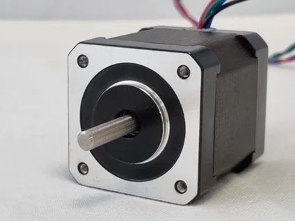
Syringe pumps use a stepper motor to deliver rotational motion, which is precisely controlled by driver electronics. Microcontrollers and advanced driver technology enable the lead screw to rotate in extremely small discrete steps, resulting in minute linear motion of the plunger and precise dispensation of small volumes of solution.

The mechanics of syringe pumps are designed to minimize frictional losses and backlash, ensuring that all rotational motion is converted to linear motion and that the torque generated by the motor is used to displace fluids rather than overcome frictional forces within the pump.

While modern syringe pumps are capable of moving very small distances, the pressure required to move viscous materials or high flow rates often necessitates a large force. If the torque generated is not high enough, the motor will stall, resulting in missing steps and failed dispensation.

Understanding the mechanics involved in the movement of the plunger and the design of the mechanical fixings and electronic control circuitry is crucial to understanding the precision movement that results in accurate and consistent dispensation of solutions.

Overall, a thorough understanding of syringe pump operation is vital to selecting appropriate specifications and ensuring accurate experimental results.


Displacement of Fluid in the Syringe


Syringe pumps operate on the principle of displacement, where the movement of the plunger results in the displacement of fluid within the syringe. The rate and volume of fluid dispensed are related to the rate and distance of plunger compression. The syringe consists of a barrel, plunger, flange, and Luer Lock for tubing connection. The volume and rate of fluid displacement can be calculated by measuring the internal diameter of the syringe and multiplying it by the distance and rate of plunger movement, respectively.

Syringe Diagram


What Moves the Syringe?


Syringe pumps use a central block that moves the plunger by converting rotational motion from a motor to linear motion using a lead screw and nut combination. Stepper motors are commonly used and controlled by driver electronics. Factors affecting syringe pump performance include the design of the central block, type of screw, connection between the screw and motor, type of stepper motor, driver electronics, and software controlling the driver. Syringe pumps are a combination of mechanical design and electronics with the stepper motor acting as the interface between the two.

Mechanics of a Syringe Pump


A syringe pump uses a lead screw mechanism to create linear motion. The lead screw is supported by a bearing at both the start and end of the moving block's travel, minimizing friction. Locking collars ensure that the screw can only rotate and not move backward or forwards. The lead screw can be connected to the motor by a coupling or drive belt, each with their own advantages and disadvantages.

A nut is used to convert the rotational motion of the lead screw into the linear motion of the moving block. The central block also has a guide in the form of a linear rail or single/dual linear rods to constrain both the carriage and the nut, preventing any rotational movement.

The distance that the central block moves for each rotation of the screw is based on the pitch of the lead screw and the number of threads. The pitch is the distance between adjacent threads. For example, a four start lead screw with a 2 mm pitch would result in 8 mm moved during each rotation of the lead screw.


Stepper Motors and Movement


A stepper motor is a type of electric motor that provides rotational movement to the lead screw, plunger, and the dispense of liquid in many machines. The motor consists of a central shaft, known as the rotor, which includes a permanent magnet and a ring of stators that are arranged around the outside of the shaft. To rotate the shaft, pairs of stators act together to attract the rotor magnet and align it with the stator. The minimum distance that the motor can rotate is referred to as a step, and in a simplified design with four pairs of stators, the minimum distance is 45 degrees.

Modern stepper motors use a hybrid design with teeth on both the rotor and stator, allowing for higher resolution of stepping. The rotor and stator teeth have equal angular spacing, and the stator pairs are offset by an angle equal to a quarter of the distance between the teeth. When the stator sets are energized, the rotor teeth align with the stator teeth, and by alternating which set is energized and which direction of current is passed through, the polarity of the stators can be in one of four different configurations.

Most stepper motors use a total of eight stators arranged in four pairs, and each stator has six teeth, resulting in an average total of 48 teeth. The rotor, however, will have 50 teeth, allowing for a stepping angle of 1.8 degrees per step. This results in a movement of 0.04 mm for an 8mm lead screw, which is larger than the minimum distance that most stepper motors can move. However, microstepping can be used to break up these steps and increase the resolution of movement.

Motor Control and Micro Stepping


A stepper motor is an electromechanical device that converts electrical pulses into rotational motion, making it ideal for precise control in many applications, including syringe pumps. The motor is controlled by a driver circuit that sends electrical pulses to the stator coils, which cause the rotor to move in a series of discrete steps. Stepper motor drivers allow for microstepping, which means that the motor can move fractions of a step by varying how energized each set of stators are. This technique can divide each individual 1.8⁰ step into as many as 256 smaller steps, resulting in an individual step angle as small as 0.007⁰.

To achieve microstepping, the driver must energize both sets of stators simultaneously and control the strength of the magnetic field generated by the stators. This is done using pulse-width modulation to reduce the average power delivered to the stators, which varies the duty cycle of the stator sets. A lower duty cycle means a lower strength magnetic field is used to move the rotor, resulting in less torque generated per microstep.

However, microstepping can lead to lower maximum force applied to the syringe, which can be problematic in some applications where high flow rates, high viscosity, or constrained flow channels are used. This is because a higher torque is required to push the syringe in these conditions, and if the torque is not high enough, the motor can stall and result in inaccurate dispense rates and volumes. Therefore, the driver must be carefully calibrated to balance the benefits of microstepping with the need for sufficient torque to achieve accurate pumping.


What are the key considerations for infusion pumps?

Portability

Infusion pumps must be portable, since patients need to be mobile both within the hospital and at home. The devices must be battery powered, relatively small, and relatively lightweight. Designers, therefore, require solutions that minimize size and power consumption. Examples include the use of switching voltage regulators instead of linear regulators, even in low-energy power supplies, and the use of higher frequency switching supplies to minimize the size of external components.

Pump Mechanism

Traditionally, stepper motors have been used in the pump mechanism to provide a precise flow rate. With angular-position sensors or Hall-effect sensors, it is possible to use DC motors instead. In these designs, the motors drive actuators (cams and fingers) to milk the tubing in precisely known fluid volumes per revolution of the mechanism.

Motor loading varies as the mechanism rotates. Motor load is affected by the position of the pump mechanism, fluid viscosity, and flow rate. To reduce power consumption, motor drive circuits can include motorload sensor signals that feed into a closed-loop control system to adjust the motor drive voltage. A variety of current-sense amplifiers, operational amplifiers, comparators, and filters are used to implement these closed-loop control systems.

Power Supplies

To maximize battery life, system designers use switch-mode voltage regulators for any significant power level. Switch-mode converters should run as fast as possible to minimize size and weight. Low-dropout linear regulators (LDOs) are used only in the very lowest power circuitry where their low efficiency can be tolerated, or where the output voltage of the LDO is not much lower than the input voltage, which keeps the efficiency high.

The use of fairly sophisticated processors places requirements on power supplies that can include voltage identification digital (VID) control from the central processing unit (CPU), fast load-step response, and precision low-voltage/high-current outputs. Digital-to-analog converters (DACs) and digital potentiometers are used in these power supplies when on-the-fly programmability is needed but VID control is not built into the regulator controller.

Because these are patient-connected devices and AC-line powered, they must meet UL® and IEC safety requirements. This means that the offline switching power supply must be designed and certified by these organizations for medical applications with patient connection.

Figure 3. Functional block diagram of an infusion pump.

Figure 3. Functional block diagram of an infusion pump.

Battery Management

Caregivers often need to transport patients while they remain on the IV, so the infusion pump must be able to operate from battery power alone for several hours. The use of rechargeable battery packs is mandatory.

The infusion pump absolutely must not run out of battery power; otherwise, it would stop pumping. Because of this, an accurate battery fuel gauge is required. Coulomb counting is the accepted method today, as voltage-sensing fuel gauges are not nearly accurate enough for this type of patient-connected equipment.

User Interface

The user interface is used to program the flow rate and provides a wealth of information. In addition to the infusion rate, hospital units display parameters such as the fluid being infused, patient information, the health of the pumping system, the amount of battery life remaining, and alarm conditions.

On some wearable models intended for home use, the patient is expected to do the programming. These devices benefit from intuitive graphical user interfaces (GUIs) that guide patients through the programming process. These infusion pumps frequently have color displays and touch screens for user inputs. Visible, audible, and haptic responses to user touch inputs help designers improve the user experience. Advanced touch-screen controllers like the MAX11811 offer haptic feedback, touch processing to reduce bus traffic, and autonomous modes for precision gesture detection.

Flow rates can be programmed over a very wide range: 0.01mL/hr to 999mL/hr is typical. Due to a history of medication errors caused by pump programming errors, sophisticated software routines have been implemented in infusion pumps to warn users when unusual or dangerous infusion rates are selected.

Displays/Keyboards

Full-color, high-resolution, backlit liquid-crystal displays (LCDs) are the most common. Some pumps also incorporate auxiliary alphanumeric displays. Display self-test at power-up is an FDA requirement, so designers require drivers with built-in self-test features.

Self-Test and System Monitoring

All infusion pumps must perform power-on self-test (POST) to meet FDA requirements. This includes tests of all critical processors, critical circuitry, indicators, displays, and alarm functionality. Some POST operations can require user observations, but additional circuitry is used for self-checking to reduce the risk of undetected failures.

For example, some models use a safety processor to monitor the performance of the main processor and to generate an alarm if unexpected behavior is detected. Another example of self-test is the simple monitoring of current through light-emitting diodes (LEDs) as they are turned on and off. If currents fall outside the acceptable range, a fault is indicated. Probably the most common self-test is the watchdog timer (WDT). Microprocessor supervisors with WDT functions are commonly used to ensure that the processor executes within proper code boundaries. In medical devices, it is usually not acceptable to have the supervisor on the same IC as the microprocessor, as this approach would subject the supervisor to the same transient errors as the microprocessor.

Supervisory functions are critical for ensuring that the pump is operating properly during patient use. Microcontrollers (of which there are often several in a single pump) must be held in reset until all power supplies are within tolerance and stable. All power supplies are monitored with voltage supervisors for undervoltage and overvoltage conditions. Motor loading is monitored and motor-stall detection is provided. (Motor stall is a critical failure causing a top-priority alarm.) Because of the criticality of the system, often power-supply voltages are monitored with ADCs so that their exact value can be recorded periodically. ADCs are also needed for sensor readings, such as temperature, motor loading, IV line pressure, and battery voltage.

Temperature sensing is implemented in the battery pack, the power supply, the motor, and the display. Due to the high efficiency of these designs, fans are usually not needed. These pumps must be splash-proof, so it is difficult to put in openings for airflow.

Alarms

Infusion pumps require audible and visible alarms to alert users to faults or potentially dangerous conditions. Bicolor or tricolor (red/orange/green) LEDs are typically used as visual indicators. Audible alarms vary from simple beepers driven by the microcontroller's pulse-width modulation (PWM) output to more sophisticated alarms (such as voice synthesis) created with an audio DAC.

Even simple audio beepers should include a self-test feature. This function can be implemented either indirectly by monitoring for a speaker impedance within range or directly by incorporating a mic near the speaker to register the audio output and confirm that it is at the proper level.

Timekeeping

Due to the criticality of patient care, every event needs to be logged and time stamped. Every key press, every start and end of an infusion, every change of configuration (pump door opening/closing, AC power disconnect, etc.), and every reported fault condition needs to be logged and time stamped for later review in case of lawsuits or instrument malfunction.

A real-time clock (RTC) is required. Since other clock sources for microprocessors and digital circuitry are not especially critical, standard crystals can be used. If extreme accuracy is needed for the RTC, Maxim has RTCs with built-in temperature-compensated crystal oscillators (TCXOs) that achieve an accuracy of ±2ppm (0°C to +40°C), which is about two orders of magnitude more accurate than a standard crystal.

Electrostatic Discharge

All infusion pumps must pass IEC 61000-4-2 electrostatic discharge (ESD) requirements by either using electronics with built-in protection or by adding ESD line protectors to exposed traces. Maxim offers many interface parts with this high ESD protection built-in, as well as stand-alone ESD diode arrays.

Interfaces

Modern infusion pumps include interfaces to connect to hospital information systems. These are variously hardwired (RS-232, RS-485, USB, and Ethernet) and/or wireless interfaces (Bluetooth® and Wi-Fi®).

For wired interfaces, galvanic isolation is critical to meet the patient safety requirements of IEC 60601-1. Interfaces with unidirectional lines (such as RS-232, RS-485, RS-422) are not difficult to isolate. The only challenge is to create an isolated supply for them residing on the isolated side. An integrated device such as the MAX256 can solve this challenge by providing up to 3W of isolated power for isolated interfaces from a compact SO package.


Tuesday, February 14, 2023

Time constant of EEG recording


  What is time Constant in EEG ?

The time constant in EEG recording refers to the time it takes for a voltage amplifier in an EEG recording system to respond to changes in the electrical potential of the brain. It determines how fast the amplifier responds to changes in the EEG signal and is a measure of the smoothing effect of the amplifier on the signal. 

The time constant is typically expressed in milliseconds and is adjustable, allowing researchers to trade off temporal resolution (the ability to capture fast changes in the signal) against noise reduction (the ability to reduce unwanted electrical interference).

A smaller time constant results in a faster response time and greater temporal resolution, but also results in more noise in the signal. 

A larger time constant results in less noise but also a slower response time and lower temporal resolution. The optimal time constant for an EEG recording depends on the research question and the characteristics of the EEG signal being recorded.

The time constant, denoted by the Greek letter tau, is a particular parameter that describes how quickly a first-order, linear time-invariant would react to a step input.

It often corresponds to the amount of time needed for a specific parameter to change by a factor of 1 1/e (approx. 0.6321).


 You may needs following basic concepts to understand Time Constant more better 

Bio-signal Filters

Biosignal information is difficult to see from the raw data. Because of this, biomedical engineers have created and used methods like relative filters and temporal (time) constants. Muscle noise, unstable dc offset, and noise distortions that may arise at the skin electrode contact can all interfere with signals. Fortunately, filters make an effort to eliminate these undesirable sounds while allowing certain signal frequencies to pass.

Bio-signal filters are digital or analog filters used to process biological signals such as electroencephalography (EEG), electrocardiography (ECG), electromyography (EMG), and others. These filters are used to enhance the quality of the signals, reduce noise, and extract relevant information from the signals. There are several types of bio-signal filters, each with its own strengths and weaknesses, including:

  1. Low-pass filters: These filters allow low-frequency signals to pass through, while blocking high-frequency signals, which are often considered noise.


  1. High-pass filters: These filters allow high-frequency signals to pass through, while blocking low-frequency signals, which are often considered to be baseline signals.

  2. Band-pass filters: These filters allow signals within a specified frequency range to pass through, while blocking signals outside of that range.

  3. Band-stop filters: These filters block signals within a specified frequency range, while allowing signals outside of that range to pass through.

  4. Notch filters: These filters are used to remove specific, interfering frequencies from the signal.

The choice of filter depends on the specific requirements of the signal and the goals of the analysis. Bio-signal filters are essential tools in many areas of biomedical research and clinical practice, allowing researchers and clinicians to gain a deeper understanding of the signals and the underlying biological processes.

Importance of filer in Clinical Medicine

Filters are used in medical devices that are used for diagnostic, testing disorders of patients. Filters in electrocardiography (ECG), electroencephalography (EEG), and electromyography (EMG) emphasize critical components by reducing extraneous noise and distortion.

Filters and temporal constants are important in recovering efficient information, assisting in achieving greater quality assurance of detection, prevention for early or beginning phases of cardiac problems, and avoiding erroneous signal readings. In a systematic process of monitoring and evaluating information, filters and temporal constants are used. It is critical to eliminate noise for  monitoring and diagnostic purposes and optimum identification of the signal for subsequent research and development.

Function of Time constant


A filter's time constant influences how quickly it responds to changes in the input signal. In other words, it governs how quickly or slowly the filter reacts to changes in the signal. The time constant is commonly represented in time units like as seconds or milliseconds, and its value controls how quickly the filter achieves steady-state output in response to a step change in the input signal.


A greater time constant often leads in a slower reaction time, which means that it takes longer for the filter to achieve its steady-state output in response to a change in the input signal.

A smaller time constant, on the other hand, leads in a faster reaction time, allowing the filter to achieve its steady-state output faster in response to a change in the input signal.

The time constant is a crucial parameter that influences the filter's overall behavior, and its value must be carefully adjusted to guarantee that the filter provides the intended results. A quick reaction time may be desirable in some applications to capture rapid changes in the input signal, whilst a longer response time may be preferable in others to eliminate noise or other undesired artifacts in the signal. The best time constant for a given application is determined by the analysis's specific objectives and aims.

Signals can be analysed more effectively if it is processed in the two domains of time and frequency, usually seen in ECGs. A step response to the step input is used in the exploration of the time response. Various processing systems that measure signals such as digital filters use time constants to aid in characterising frequency responses to these filters. These responses are used to appropriate which models and methods can be applied to first order LTI systems.

The time constant τ is related to the cutoff frequency fc, an alternative parameter of the RC circuit, by

τ = R C = 1/ 2 π f c

Where time constant (in seconds) of an RC circuit, is equal to the product of the circuit resistance (in ohms) and the circuit capacitance (in farads), i.e.


Search This Blog